 Epistemis
Real clinical images, not illustrations
6,593 images and sounds across 15 collections, each with its provenance and licence in plain sight. Neither the exam nor the ward will show you a drawing.
- 6,593images and sounds
- 15collections
- 38auscultation sounds
- 100%with declared provenance
What is there exactly, collection by collection
Anyone can write "thousands of images". This is the real split of the publishable catalogue Epistemis delivers by module.
| Collection | Items | What it covers |
|---|---|---|
| Electrocardiograms | 4,099 | Real tracings with a systematic read |
| Radiology | 1,214 | X-ray, CT and MRI with findings marked up |
| Histology | 325 | Slides with their stain and magnification |
| Pathology | 263 | Gross and micro with reasoned diagnosis |
| Dermatology | 161 | Elementary lesions and classic presentations |
| Anatomy | 99 | Structures with relations and landmarks |
| Physiology | 76 | Curves and schematics of the mechanisms |
| Biochemistry | 62 | Metabolic pathways and their control points |
| Microbiology | 61 | Cultures, stains and morphology |
| Immunology | 44 | Responses and cascades |
| Fundus | 43 | Retinal images with signs marked |
| Pharmacology | 42 | Illustrated mechanisms of action |
| Embryology | 42 | Development and associated malformations |
| Auscultation | 38 | Real heart and lung sounds |
| Blood smears | 24 | Peripheral blood and marrow |
Seeing an image is not recognising one
Covered first, explained after
Every item opens with the findings hidden so you can read it yourself, then reveals them. Recognising with the answer already on screen trains nothing.
The sounds, with headphones
38 cardiac and pulmonary auscultation recordings. You do not learn a murmur by reading its description.
With licence and provenance
Every image declares where it comes from and under which licence it is used. That is what separates an atlas from a folder of screenshots.
Downloads by module
Images download when you open them or install their module, then remain in the device cache for later use with no signal. You can remove a module to recover space.
The body by systems, regions and layers
414 interactive scenes organise 7,551 anatomical structure appearances into 55 downloadable packs. The compositor overlays muscles, arteries, veins, nerves, organs, and bones in the same space, with a semitransparent guide model for following course and depth.
- 4143D scenes
- 7,551structure appearances
- 55on-demand packs
- 8body regions
- Bones, joints, muscles, fasciae, tendons and ligaments.
- Arteries, veins, lymphatics, brain, nerves and sensory organs.
- Respiratory, digestive, urinary, reproductive and endocrine systems.
- Composable layers with independent opacity, selection, and a record for every overlaid structure.
- Automatic skeletal reference that orients the region without hiding the course being studied.
- Models downloadable by region or system, with offline caching after the first download.
To study with, not to diagnose with
Epistemis is educational and revision material. It is not a medical device, it does not diagnose or prescribe, and it is no substitute for formal medical training, current clinical guidelines or a health professional's judgement.
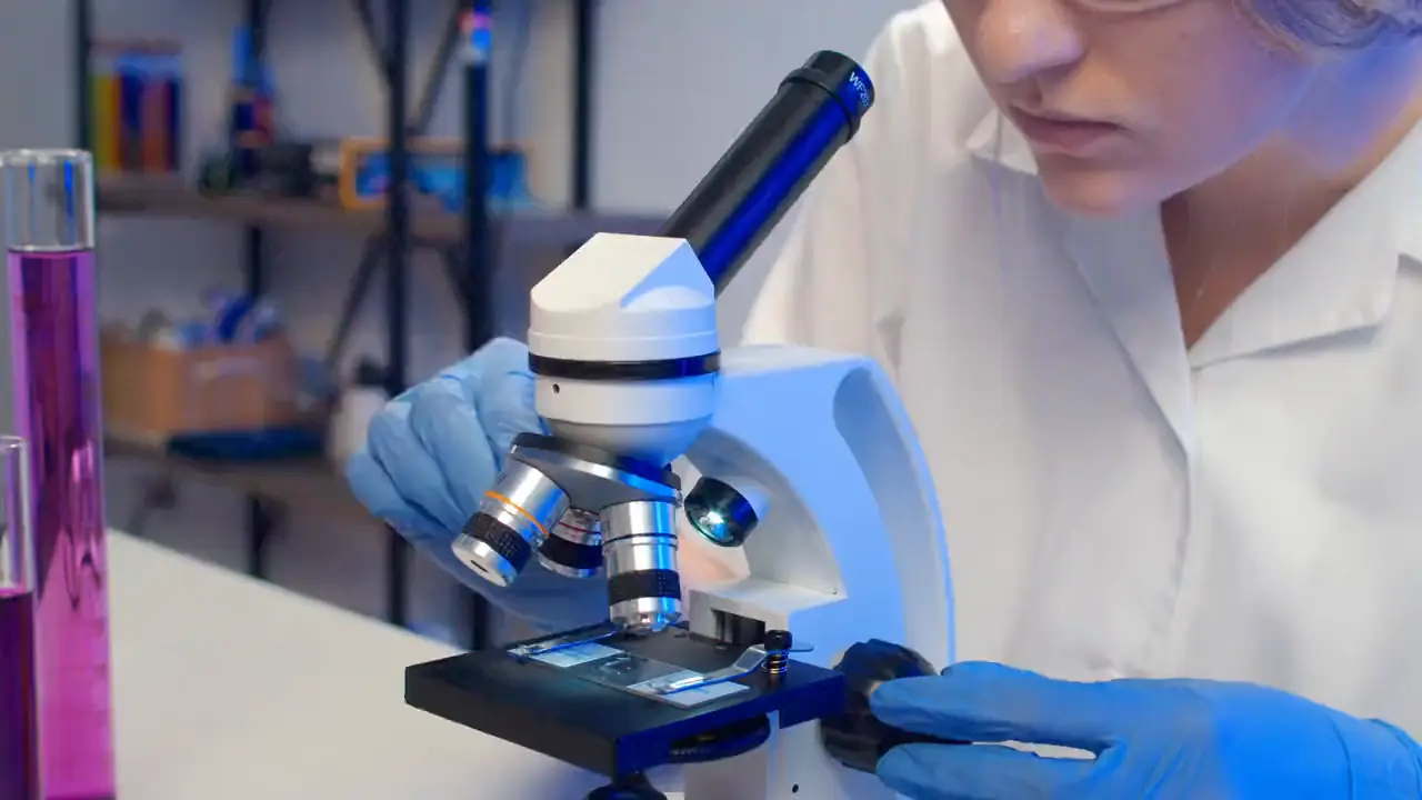
Real specimens,
with their provenance
Not a single atlas image is a stock illustration. They come from reviewed public banks, and every card carries its source and licence in writing — even when that looks ugly.
What sits behind every plate
A light box, a microscope and somebody revising. The atlas comes out of the same places where the studying happens.
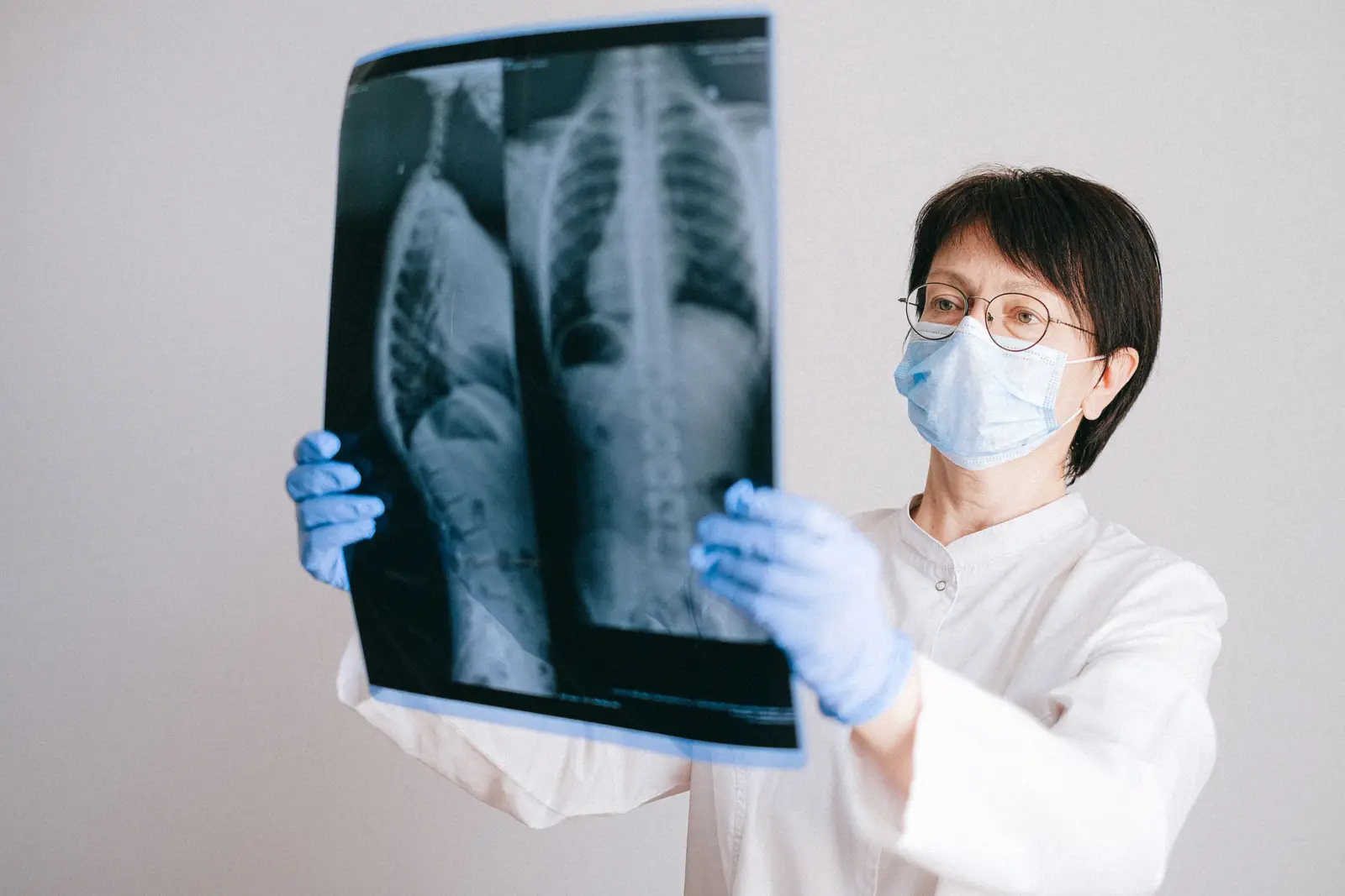
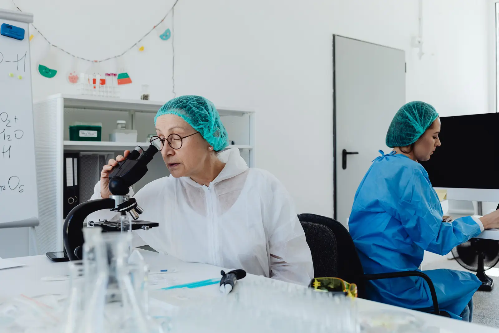
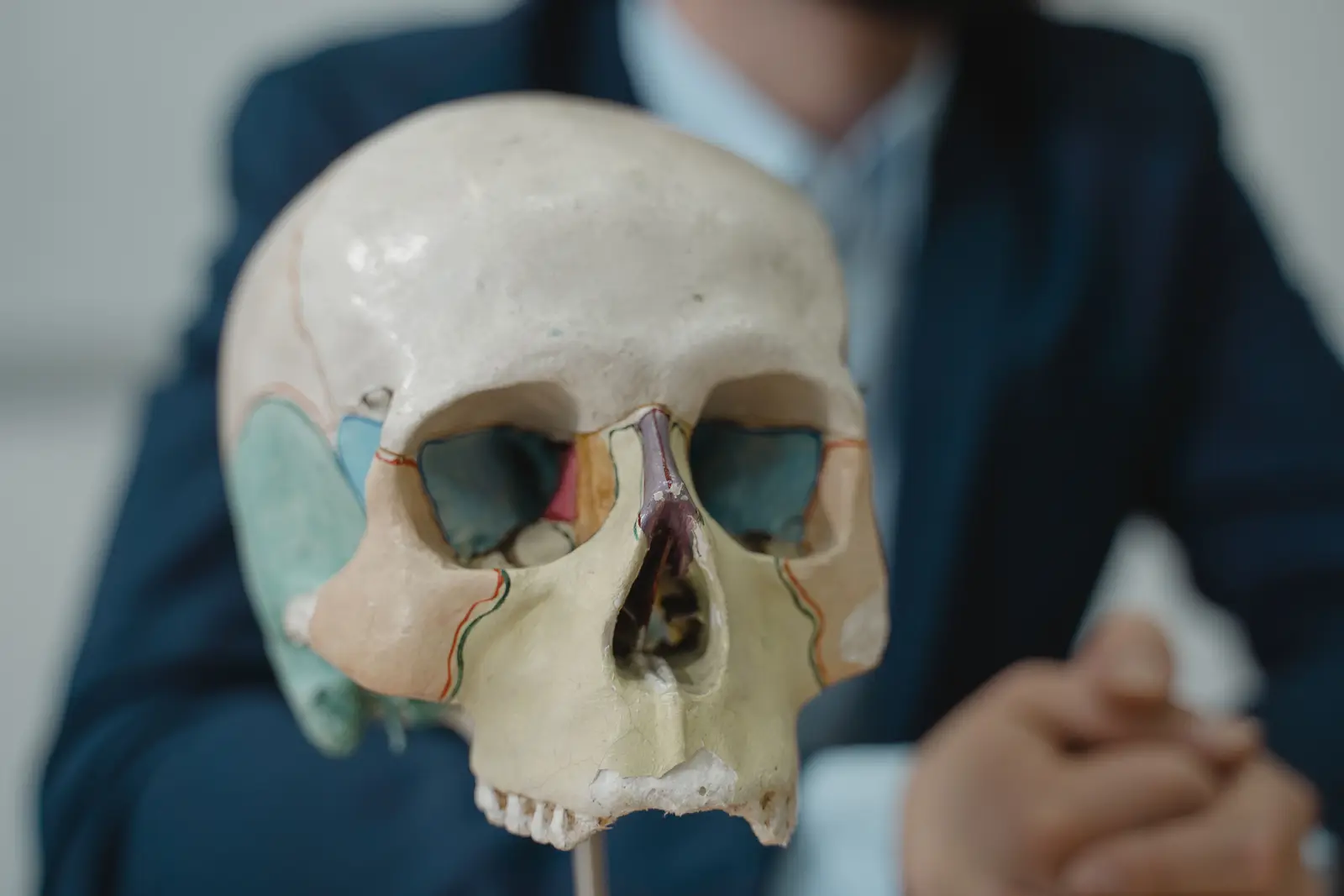

An atlas that prints the diagnosis next to the image does not train you to look: it trains you to read the caption.
Download only what you study
No account is required. Opening an image or model for the first time needs the internet; it then remains available offline while it stays cached.
Free. No account required. Offline core; the Atlas downloads by module.