 Epistemis
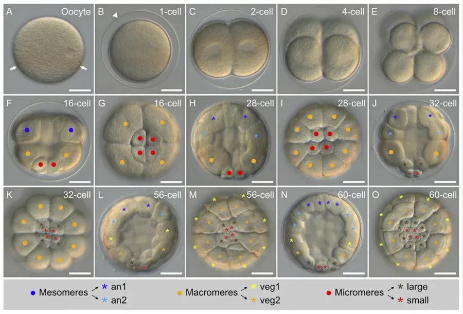
Zygote segmentation
Embryology · First weeks
The guided interpretation is available in the app
Epistemis explains the visible findings, their rationale, and important mimics through guided interpretation and clinical pearls. Free and available offline.
Epistemis is educational review material. It is not a medical device, does not diagnose or prescribe treatment, and does not replace formal medical training, current clinical guidelines, or professional clinical judgment.
- Category
- First weeks
- Difficulty
- media
- Type
- Diagram
Origin: Laurent Formery, Axel Wakefield, Maeva Gesson, Ludovic Toisoul, Guy Lhomond, Laurent Gilletta, Régis Lasbleiz, Michael S · CC BY 4.0 · Wikimedia Commons · License CC BY 4.0
Download Epistemis